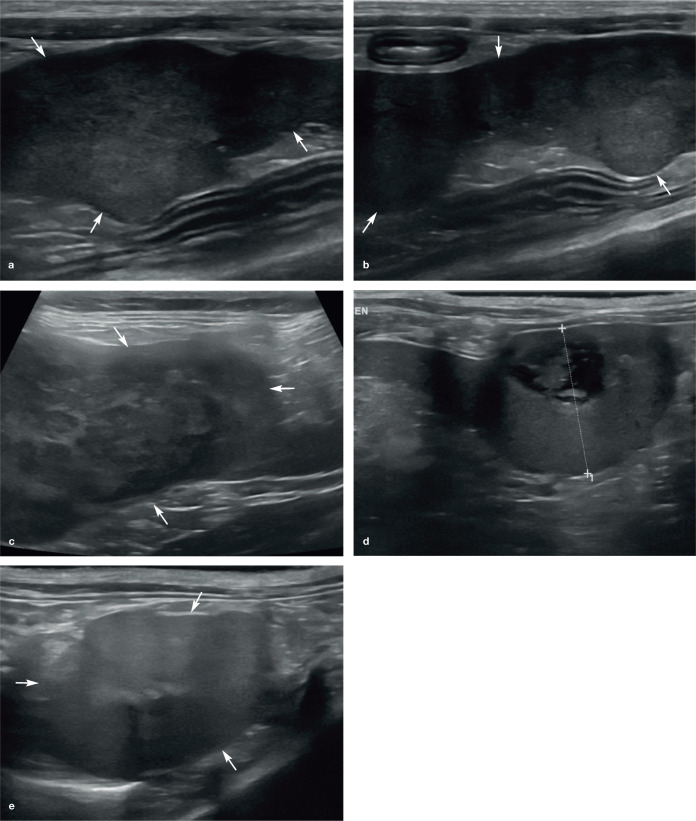
Figure 10.
Splenic masses. (a,b) Multiple large masses (arrows) are present within the diffusely hypoechoic spleen of a 10-year-old neutered female domestic shorthair cat due to metastatic mast cell neoplasia. (c) Heterogeneous splenic mass (arrows) in an 11-year-old neutered male domestic shorthair cat with lymphoma. Marked intra-abdominal lymphadenopathy and a small peritoneal effusion were also identified during the ultrasound examination. (d) Large, well-circumscribed mass (between the measuring calipers) at the tail of the spleen in a 12-year-old neutered female domestic longhair cat with carcinomatosis. The mass is isoechoic relative to the remaining splenic parenchyma with an eccentrically located heterogeneously anechoic region. The spleen was normal on ultrasound examination performed 3 months earlier and metastatic or possibly primary neoplastic splenic disease were considered the most likely differentials. (e) A large, approximately 3 cm diameter, well-defined mass (arrows) is visible at the tail of the spleen of an 11-year-old neutered female domestic shorthair cat. There is a centrally located focal hyperechoic region within the mass associated with distal acoustic shadowing. Results from ultrasound-guided fine-needle aspiration were consistent with nodular lymphoid hyperplasia

