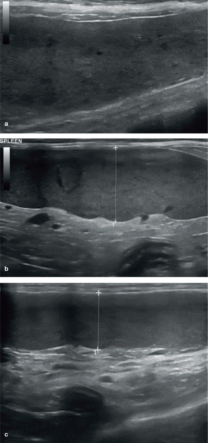
Figure 7.

(a–c) Diffuse splenomegaly (indicated between the measuring calipers in [b] and [c]) in three cats owing to lymphoma. In (a) and (b), the splenic parenchyma is heterogeneous due to the presence of small, often coalescing, hypoechoic foci; in (c), the parenchymal echotexture is uniform. In all three cases there was concurrent hepatomegaly and abdominal lymphadenopathy. A video showing splenic lymphoma is available as supplementary material
