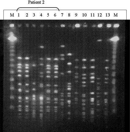
FIG. 1.
PFGE patterns produced by SmaI macrorestriction from clinical isolates reported as CoNS and Kocuria spp. by Vitek 2. Lanes 1 to 6 contain isolates from patient 2, reported as Kocuria (lanes 1, 2, and 4) and CoNS (lanes 3, 5, and 6). The isolates identified as K. varians from the pacemaker pocket (lane 1) and an electrode (lane 2) were identical to each other, as well as to CoNS isolates from another electrode and blood (lanes 5 and 6, respectively). Another CoNS cultured from blood (lane 3) and an isolate identified as K. varians that was cultured from an electrode (lane 4) were of different clones. Lanes 7 to 13 each contain an isolate identified as Kocuria spp. from a different patient, shown by PFGE to belong to different clones. Lanes M, lambda standard markers. Isolate 7 was identified by 16S rRNA gene sequencing as S. haemolyticus; all other isolates were identified as S. epidermidis.

