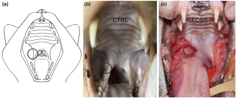
Figure 1.
(a) Image representing the feline oral cavity. The circle represents the area from which the oral mucosa samples were collected. (b) Photograph showing the oral cavity of a 4-year-old intact male domestic shorthair (DSH) control cat with no evident lesions of the mucosa (post mortem). (c) Photograph showing the oral cavity of a 6-year-old neutered female DSH cat affected by feline chronic gingivostomatitis (FCGS) exhibiting marked inflammation, ulceration and hyperplasia of the caudal oral mucosa

