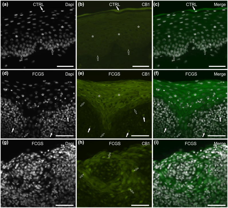
Figure 2.
Photomicrographs of cryosections of feline caudal oral mucosa showing cannabinoid receptor 1 (CB1R) immunoreactivity (IR) in tissues of the (a–c) control cats (CTRL) and (d–i) the cats affected by feline chronic gingivostomatitis (FCGS). (a–c) The white arrow indicates a 4′,6-diamidino-2-phenylindole (DAPI)-labelled nucleus of a superficial epithelial cell, displaying bright cytoplasmic CB1 immunoreactivity (IR); the stars indicate the DAPI-labelled nuclei of the epithelial cells of the intermediate layers, showing moderate cytoplasmatic CB1R IR; the open arrows indicate the DAPI-labelled nuclei of the basal layer cells, which were CB1R negative. (d–f) The stars represent the DAPI-labelled nuclei of the epithelial cells, displaying bright cytoplasmatic CB1R IR (intermediate layers); the open arrows indicate the DAPI-labelled nuclei of the basal layer cells, which were CB1R negative; the white arrows indicate the DAPI-labelled nuclei of the immunocytes localised in the lamina propria/submucosa, showing bright membrane CB1R IR. (g–i) The open arrows indicate the DAPI-labelled nuclei of the epithelial cells with a round organisation, expressing moderate cytoplasmic and bright nuclear CB1R IR. Bar = 50 µm

