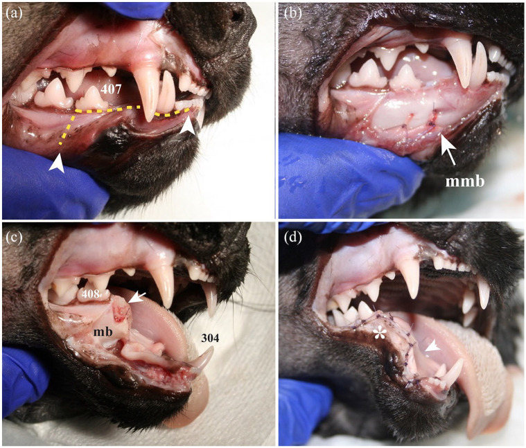
Figure 6.
Lateral view of the rostral aspect of the right mandible of a cat head specimen showing the main features of the rostral segmental mandibulectomy. (a) A surgical marker pen was used to outline the planned incisions along the gingival sulcus of the mandibular incisor teeth and labial/buccal mucosa (arrowheads). (b) A full-thickness mucoperiosteal incision made with a number 15 scalpel blade and sharp dissection with a periosteal elevator allowed exposure of the mandibular body and placement of a ligature around the middle mental neurovascular bundle with 4-0 absorbable monofilament suture material (mmb). (c) After removing the rostral aspect of the mandible and placing the ligature around the inferior alveolar neurovascular bundle, the distal root of the right mandibular third premolar tooth was extracted. Sharp alveolar bone edges (arrow) and the remaining portion of the mandibular body (mb) were smoothed with a #22 round diamond bur. (d) The labial/buccal flap was sutured over the wound, using a 5-0 absorbable monofilament suture material in a simple interrupted pattern. The remaining mandibular body (asterisk) provoked tension on the mucoperiosteal and flap (arrowhead), resulting in a decreased vertical range of motion of the contralateral mandible postoperatively.
407 = right mandibular third premolar; 408 = right mandibular fourth premolar; 304 = left mandibular canine teeth

