FIG. 1.
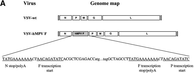
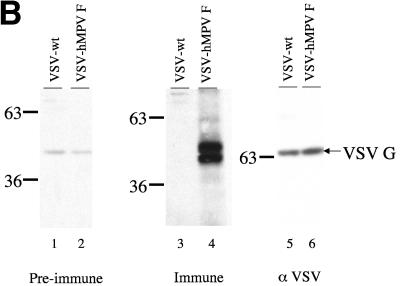
(A) Cloning and expression of the hMPV F gene in recombinant VSV. The hMPV F gene from a clinical specimen was amplified by RT-PCR and cloned into a plasmid containing the full-length genome of VSV. Recombinant VSV with the hMPV F gene inserted between the VSV N and P genes (VSV-hMPV F) was recovered by using reverse genetics. The genomic maps (3′ to 5′ orientation for the negative-strand RNA genome) of VSV-wt and VSV-hMPV are displayed in panel A. Sequences of the gene junctions (corresponding the positive-sense cDNA) are shown below the genomic maps. The transcription start and stop/poly A signals are indicated. The hMPV initiation codon (atg) and termination codon (tag) and the obligatory intergenic CT are shown. (B) Western blot of VSV-wt and VSV-hMPV F lysates with rabbit anti-hMPV F peptide serum. Proteins of infected cells were separated by polyacrylamide gel electrophoresis under reducing, denaturing conditions, transferred to nitrocellulose, and blotted with the rabbit anti-hMPV F peptide antiserum (preimmune or immune) or rabbit anti-VSV antiserum. Antibody binding was detected following incubation with a horseradish peroxidase-conjugated anti-rabbit antibody and enhanced chemiluminescence. Lane 1, VSV-wt and preimmune serum; lane 2, VSV-hMPV F and preimmune serum; lane 3, VSV-wt and immune serum; lane 4, VSV-hMPV F and immune serum; lane 5, VSV-wt and rabbit anti-VSV serum; lane 6, VSV-hMPV F and anti-VSV serum. The positions of the molecular weight markers for each panel are indicated.


