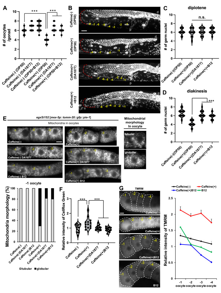
Figure 3.
Effects of vitamin B12 on oogenesis in CIA. (A) Quantification of the number of oocytes produced by CIA fed with OP50 or DA1877 with or without vitamin B12. (B–D) Representative gonad images of DAPI staining (B) show the number of germ nuclei in diplotene (C) and diakinesis (D) in CIA fed the corresponding bacterial diet with vitamin B12 supplementation. The red dotted line indicates diplotene. Bar, 20 μm. The yellow-colored number indicates the developing oocytes that aligned from the proximal region; d, distal side of the gonad arm; p, proximal side of the gonad arm. (E) Comparison of mitochondrial morphology in the oocytes of caffeine-ingested EGD623 (egxSi152 [mex-5p: tomm-20::gfp::pie-1]) transgenic animals fed OP50 or DA1877 and supplemented with or without vitamin B12. The type of mitochondrial morphology was classified as tubular or globular. Bar, 5 μm. (F,G) Comparison of mitochondrial reactive oxygen species through CellROX Green staining (F) and mitochondrial membrane potential through TMRM staining (G) in the oocytes of CIA fed with OP50 or DA1877 supplemented with or without vitamin B12. Bar, 5 μm. The violin plot shows the relative level of intensity of CellRox Green (F). The line graph shows the relative intensity of TMRM in oocytes. Data represent mean ± standard deviation. n.s., p > 0.05. ***, p < 0.001. Number of analyzed animals, n ≥ 20 in respective conditions.

