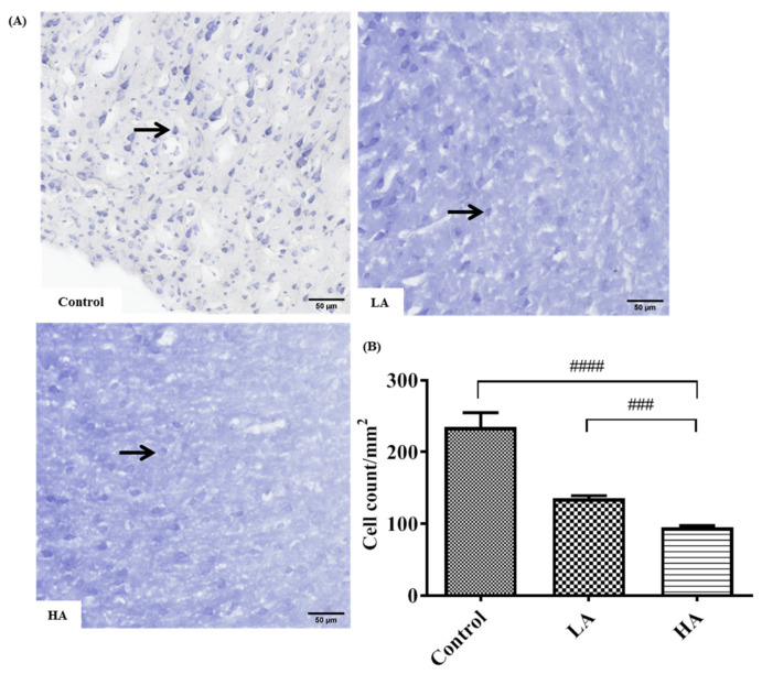
Figure 3.
Effects of aspartame oral administration on the Nissl stain of the rats’ cerebral cortex (20×). (A) Control group shows deep Nissl staining in pyramidal cells. Low dose of aspartame (LA) shows mild Nissl staining in pyramidal cells. High dose of aspartame (HA) show poor Nissl staining, with least number of positive Nissl-stained pyramidal cells. (B) Population of Nissl-stained neurons in the cerebral cortex. Data are shown as mean ± SD; ### p < 0.001, #### p < 0.0001 when compared with control group. Black arrow refers to positive Nissl-stained pyramidal cells.

