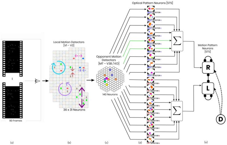
Figure 1.
Schematic of the model in one hypothetical point in time, from left to right: (a) the reel of biological motion stimulus; (b) local motion detectors as ensemble of 1116 neurons positioned in a 36 by 31 arrangement, firing due to the motions they have experienced during two consecutive frames, are represented by the cells with color−filled arrows (blue: right, orange: left, grey: up, green: down); the larger, two−headed or curved, colorful arrows were drawn to display the types of opponent motions that would be sensed on the next level (cyan: horizontal expansion, orange: vertical expansion, magenta: vertical contraction, green: counter−clockwise rotation, and yellow: clockwise rotation); (c) opponent motion detectors, the ensemble of 140 neurons to detect horizontal expansion, horizontal contraction, vertical expansion, and vertical contraction, the activated detectors are marked with color−filled hexagons with their corresponding color (cyan: horizontal expansion, orange: vertical expansion, magenta: vertical contraction, green: counter−clockwise rotation, and yellow: clockwise rotation); (d) optical−flow pattern detectors, an arrangement of 18 neurons following a one−dimensional mean−field dynamics, each neuron incorporates a statistical template (displayed as colorful map) that represents a specific part of the manifold of the kicking sequences (for example neuron number 2 contains a template for the seconds 11 to 20 of the kick−to−right sequence, while neuron number 10 would have a larger instantaneous input for the seconds 1 to 10 of the kick to the left stimulus). Green arrows highlight the contribution of two cells to the evidence integration at that hypothetical point due to the similarity of the evidence signal and their template (e) thresholding stage, two decision neurons for the right and left decisions (marked by capital letters R and L on the square cells with soft edges) are follow our mutual inhibition dynamics, receiving their corresponding inputs from integration stage, and the straight and curve lines with rounded heads highlight the inhibitory interaction between the neurons and the auto-inhibition, respectively. No activity could be seen by either of the neurons since at that hypothetical point in time, neither had made a decision yet. Also, the dotted curved arrows and the circle with the letter D represent the disremembering mechanism.

