Abstract
A case of non-catheter-related bacteremia caused by Chryseobacterium indologenes in a nonneutropenic man with a solid tumor is described. The patient was successfully treated with piperacillin-tazobactam.
CASE REPORT
A man, aged 54, with squamous cell carcinoma of the right nasal tube and multiple metastases in the regional cervical lymph nodes was admitted in May 2004 to the Metaxa Anticancer Hospital because of severe mass hemorrhage and the inability to swallow. In October 2003 the primary lesion and the regional lymph nodes were excised in another hospital. Between these two hospital visits, the patient had received chemotherapy and regional radiotherapy. Because of the severe hemorrhage, regional hemostatic radiotherapy was administered and the patient was fed through a gastrostomy. In addition, he received chemotherapy (methotrexate and gemcitabine) on a weekly basis for a month. The patient remained in stable condition.
On day 46, while in remission with a normal white blood cell count (7 × 10⁹ cells/liter), the patient developed rigors and a temperature of 39°C. No source of infection was clinically apparent. Laboratory investigation revealed a white blood cell count of 22 × 109/liter (84% granulocytes, 9% lymphocytes, and 7% monocytes), a hemoglobin level of 7.9 g/dl, a platelet count of 116 × 109/liter, a γ-glutamyl transpeptidase level of 215 IU/liter, and an alkaline phosphatase level of 169 IU/liter. All of the remaining biochemical laboratory tests were unremarkable. Chest X ray as well as a computed tomography scan of the brain, chest, and abdomen showed no findings of infection. Urine, wound, and blood cultures obtained through a Hickman catheter were negative. However, blood cultures obtained through a peripheral venous site grew a gram-negative rod, later identified as Chryseobacterium indologenes. The patient was treated with piperacillin-tazobactam (4.5 g every 8 h intravenously) for 10 days. The fever resolved 4 days later. There was no evidence of relapse during the next month, when he was transferred to another hospital. After identification of the microorganism, the catheter was removed, but the semiquantitative culture of the tip was negative. Environmental samples, e.g., sink, showerhead, taps, detergents, and disinfectants, from the patient's medical ward were collected and cultured as described by Perola et al. (14). All environmental samples failed to yield C. indologenes.
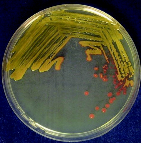
Three sets of standard medium blood cultures in a total of six bottles were drawn within 24 h. The patient was not receiving antimicrobial agents at the time of collection. Cultures were processed with the BacT/ALERT System (BioMerieux, Marcy l'Etoile, France). Bacterial growth was detected within 24 to 48 h. The three aerobic bottles grew smooth, circular, yellow-pigmented colonies on sheep blood agar. Yellow-pigmented colonies were also observed on peptone medium. The flexirubin type of pigment was confirmed by adding 1 drop of 10% KOH solution to a bit of cell paste. The color of the colonies changed from yellow to red (Fig. 1). The organism was identified as C. indologenes by both conventional biochemical reactions, as previously described (18), and the API 20NE (biotype 2650205; probability, 99.9%) identification system (BioMerieux). The identification was confirmed by an independent laboratory using the Vitek ID-GNB (BioMerieux) identification system (typicity index [0.28] and tests contraindicating typical biopattern BGLU [beta-glucosidase][0.05] and BNAG [b-n-acetyl-glucosaminidase][0.16]).
FIG. 1.
Yellow-pigmented colonies of C. indologenes in peptone medium. The yellow color, due to the production of flexirubin, turns to red after the culture is poured on 10% KOH solution.
Antibiotic sensitivity testing was performed using the panel Neg MIC type 30 (Micro Scan; Dade Behring, Inc., West Sacramento, Calif.). Control strains were Escherichia coli ATCC 25922 and Pseudomonas aeruginosa ATCC 27853. The sensitivity of the isolate to the antimicrobial agents was determined by applying the NCCLS susceptibility criteria used for P. aeruginosa (11). The MICs for the isolate were as follows: ≤ 0.5 mg/liter for ciprofloxacin, ≤ 2 mg/liter for levofloxacin, ≤ 8 mg/liter for piperacillin-tazobactam, and ≤ 2/38 mg/liter for co-trimoxazole. The isolate was resistant to all other antibiotics tested, such as amikacin, gentamicin, tobramycin, piperacillin, cefepime, cefotaxime, ceftazidime, aztreonam, meropenem, imipenem, tetracycline, and chloramphenicol.
Chryseobacteria are a group of nonmotile, catalase-positive, oxidase-positive, indole-positive, non-glucose-fermenting, gram-negative bacilli. The genus Chryseobacterium includes six species that were previously designated members of the genus Flavobacterium (17). Chryseobacterium gleum and Chryseobacterium indologenes, previously known as Flavobacterium CDC group IIb, have been clearly differentiated by DNA-DNA homology and eight phenotypic characteristics (18). Although Chryseobacterium meningosepticum is the most pathogenic member of the genus, C. indologenes is the species most commonly reported to cause different clinical syndromes usually associated with various indwelling devices (6, 16).
Chryseobacteria are not a part of the human flora but are found in soil, plants, and foodstuffs (16). In the hospital environment, these organisms exist in water systems and on wet surfaces of medical tools and equipment (10, 16). Chryseobacteria represent only 0.03% of all bacterial isolates collected by the SENTRY Program during the period 1997 to 2001, and they are responsible for 0.03% of all bloodstream infections (8). Chryseobacteria are of low pathogenicity. The production of biofilm on foreign materials and protease activity may play an important role in the virulence of invasive infections due to C. indologenes (5, 13).
Infections by C. indologenes affect mainly patients from Taiwan. To date, 38 cases have been referred in that country (6, 9, 10). Conversely, only two cases in Australia (7), one case in the United States (4), and three cases in Europe (2, 12, 15) have been described. Intra-abdominal infections, primary or catheter-related bacteremia, and wound sepsis are the most common clinical syndromes in Taiwan, while malignancies and diabetes mellitus are the main underlying diseases. Approximately half of the patients developed nosocomial infections associated with various indwelling devices, whereas three of the five (14%) fatal cases involved polymicrobial infections. Therapy does not usually require removal of an indwelling device (6).
The choice of an effective drug for empirical treatment of infections due to Chryseobacterium spp. is sometimes difficult. According to the results of the SENTRY Antimicrobial Surveillance Program, the most active agents against C. indologenes are the quinolones (garenoxacin, gatifloxacin, and levofloxacin) and trimethoprim-sulfamethoxazole (≥95% susceptibility), followed by piperacillin-tazobactam (90% susceptibility). Ciprofloxacin, cefepime, ceftazidime, piperacillin, and rifampin showed reasonable activity (85% susceptibility). On the contrary, aminoglycosides, other β-lactams, chloramphenicol, linezolid, and glycopeptides are not appropriate for treating infections due to this organism (8). Clinical microbiologists have two additional problems to resolve: (i) susceptibility disk diffusion testing is inaccurate for chryseobacteria (1, 3) and (ii) the MIC breakpoints for these organisms have not been established by the NCCLS (11).
In conclusion, this case shows that C. indologenes should be added to the list of agents that cause severe infections. Although the majority of C. indologenes infections are linked to the use of indwelling devices during a hospital stay, non-catheter-related bacteremia may also occur. In these patients, wound infections and cellulitis are the most common portals of entry of the organism into the circulation. Although C. indologenes is widely distributed in hospital environments, the source of infection in the majority of infections remains unknown. It is likely that the establishment of an infection requires the presence of the following factors: the production of a biofilm on foreign materials, a suitable portal of entry, and immunodeficiency. When significant infections due to C. indologenes are encountered, susceptibility testing by a broth microdilution technique is necessary. Piperacillin-tazobactam may appear to be promising for treatment of severe infections due to this organism.
Acknowledgments
We thank Catherine Ioannou (BioMerieux) for her laboratory assistance.
REFERENCES
- 1.Aber, R. C., C. Wennersten, and R. C. Moellering, Jr. 1978. Antimicrobial susceptibility of flavobacteria. Antimicrob. Agents Chemother. 14:483-487. [DOI] [PMC free article] [PubMed] [Google Scholar]
- 2.Doiz, O., M. T. Llorente, A. Mateo, C. Seral, C. Garcia, and M. C. Rubio. 1999. Corneal abscess by Flavobacterium indologenes. Enferm. Infecc. Microbiol. Clin. 17:149-150. [PubMed] [Google Scholar]
- 3.Fraser, S. L., and J. H. Jorgensen. 1997. Reappraisal of the antimicrobial susceptibilities of Chryseobacterium and Flavobacterium species and methods for reliable susceptibility testing. Antimicrob. Agents Chemother. 41:2738-2741. [DOI] [PMC free article] [PubMed] [Google Scholar]
- 4.Green, B. T., and N. E. Nolan. 2001. Cellulitis and bacteremia due to Chryseobacterium indologenes. J. Infect. 42:219-220. [DOI] [PubMed] [Google Scholar]
- 5.Hsueh, P. R., L. J. Teng, S. W. Ho, W. C. Hsieh, and K. T. Luh. 1996. Clinical and microbiological characteristics of Flavobacterium indologenes infections associated with indwelling devices. J. Clin. Microbiol. 34:1908-1913. [DOI] [PMC free article] [PubMed] [Google Scholar]
- 6.Hsueh, P. R., L. J. Teng, P. C. Yang, S. W. Ho, W. C. Hsieh, and K. T. Luh. 1997. Increased incidence of nosocomial Chryseobacterium indologenes infections in Taiwan. Eur. J. Clin. Microbiol. Infect. Dis. 16:568-574. [DOI] [PubMed] [Google Scholar]
- 7.Kienzle, N., M. Muller, and S. Pegg. 2001. Chryseobacterium in burn wounds. Burns 27:179-182. [DOI] [PubMed] [Google Scholar]
- 8.Kirby, J. T., H. S. Sader, T. R. Walsh, and R. N. Jones. 2004. Antimicrobial susceptibility and epidemiology of a worldwide collection of Chryseobacterium spp.: report from the SENTRY Antimicrobial Surveillance Program (1997-2001). J. Clin. Microbiol. 42:445-448. [DOI] [PMC free article] [PubMed] [Google Scholar]
- 9.Lin, J. T., W. S. Wang, C. C. Yen, J. H. Liu, T. J. Chiou, M. H. Yang, T. C. Chao, and P. M. Chen. 2003. Chryseobacterium indologenes bacteremia in a bone marrow transplant recipient with chronic graft-versus-host disease. Scan. J. Infect. Dis. 35:882-883. [DOI] [PubMed] [Google Scholar]
- 10.Lu, P. C., and J. C. Chan. 1997. Flavobacterium indologenes keratitis. Ophthalmologica 211:98-100. [DOI] [PubMed] [Google Scholar]
- 11.NCCLS. 2000. Methods for dilution antimicrobial susceptibility tests for bacteria that grow aerobically. Approved standard M7-A4. NCCLS, Wayne, Pa.
- 12.Nulens, E., B. Bussels, A. Bols, B. Gordts, and H. W. Van Landuyt. 2001. Recurrent bacteremia by Chryseobacterium indologenes in an oncology patient with a totally implanted intravascular device. Clin. Microbiol. Infect. 7:391-393. [DOI] [PubMed] [Google Scholar]
- 13.Pan, H. J., L. J. Teng, Y. C. Chen, P. R. Hsueh, P. C. Yang, S. W. Ho, and K. T. Luh. 2000. High protease activity of Chryseobacterium indologenes isolates associated with invasive infection. J. Microbiol. Immunol. Infect. 33:223-226. [PubMed] [Google Scholar]
- 14.Perola, O., T. Nousiainen, S. Suomalainen, S. Aukee, U.-M. Karkkainen, J. Kauppinen, T. Ojanen, and M.-L. Katila. 2002. Recurrent Sphingomonas paucimobilis-bacteremia associated with a multi-bacterial water-borne epidemic neutropenic patients. J. Hosp. Infect. 50:196-201. [DOI] [PubMed] [Google Scholar]
- 15.Sanchez, M. F., P. R. Diaz, J. F. Ucelay, and D. A. Perez-Navaro. 1993. Pneumonia from Flavobacterium indologenes (group II B) in an immunodepressed patient. An. Med. Interna 10:312. [PubMed] [Google Scholar]
- 16.Schreckenberger, P. C., M. I. Daneshvar, R. S. Weyant, and D. G. Hollis. 2003. Acinetobacter, Achromobacter, Chryseobacterium, Moraxella, and other nonfermentative gram-negative rods, p. 749-779. In P. R. Murray, E. J. Baron, J. H. Jorgensen, M. A. Pfaller, and R. H. Yolken (ed.), Manual of clinical microbiology, 8th ed. American Society for Microbiology, Washington, D.C.
- 17.Vandamme, P., J. F. Bernardet, P. Segers, K. Kersters, and B. Holmes. 1994. New perspectives in the classification of the flavobacteria: description of Chryseobacterium gen. nov., Bergeyella gen. nov., and Empedobacter nom. rev. Int. J. Syst. Bacteriol. 44:827-831. [Google Scholar]
- 18.Yabuuchi, E., Y. Hashimoto, Y. Ezaki, Y. Ido, and N. Takeuchi. 1990. Genotypic and phenotypic differentiation of Flavobacterium indologenes Yabuuchi et al. 1983 from Flavobacterium gleum Holmes et al. 1984. Microbiol. Immunol. 34:73-76. [DOI] [PubMed] [Google Scholar]


