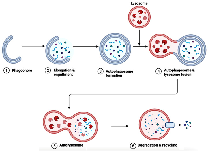
Figure 6.
A diagram depicting the steps in autophagy. The elongation of phagophores begins when LC3 attaches to the membrane. (1) The phagophore expands when the cytoplasmic materials (e.g., mitochondria, endoplasmic reticulum, aggregated proteins, bacteria, and virus) are engulfed. (2) Then, the phagophore undergoes both elongation and engulfment. (3) This creates the structure called an autophagosome. (4) Then, the lysosome fuses with the autophagosome. (5) The fusion forms the autolysosome. (6) Lysosomal hydrolases cause degradation and recycling cellular faulty or damaged components. The process of autophagy is crucial for several functions in the body, such as cell survival, degrading protein aggregates, and homeostasis. Autophagy is impaired in AD, blocking the degradation and recycling of faulty proteins and other cellular components. This figure was created with BioRender.com.

