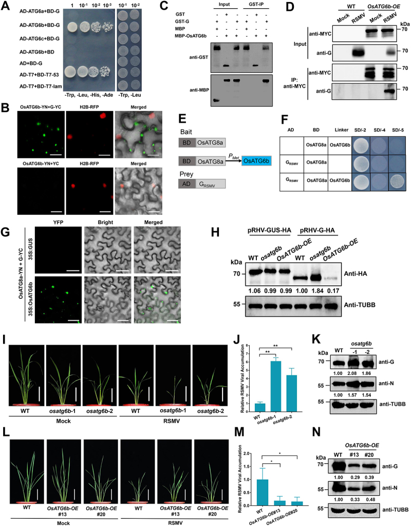
Figure 6.

OsATG6b targets RSMV glycoprotein and restricts RSMV infection. (A) Y2H assay showing the interaction between OsATG6b and RSMV glycoprotein. Yeast strain Y2HGold cells co-transformed with the indicated plasmids were cultured separately on the SD-Trp-Leu-His-Ade and SD-Trp-Leu selection medium. (B) BiFC analysis of the interaction between OsATG6b and RSMV glycoprotein. OsATG6b-YN was coexpressed with G-YC or YC in RFP-H2B transgenic N. benthamiana leaves. YFP signals were visualized by confocal microscopy. The nuclei showed red fluorescence. Bars: 20 μm. (C) GST affinity-isolation assay showing the interaction between OsATG6b and RSMV glycoprotein in vitro. Purified MBP-OsATG6b or MBP was incubated with GST-G. After being immunoprecipitated with glutathione-Sepharose beads, the proteins were detected by immunoblot with anti-MBP or anti-GST antibodies. (D) co-IP analysis of the interaction between OsATG6b and RSMV glycoprotein. WT and OsATG6b-OE plants were mock or RSMV inoculated and were harvested at 15 dpi. Total proteins were immunoprecipitated with anti-MYC beads. Input and IP proteins were analyzed by immunoblot with anti-MYC and anti-G antibodies. (E) Schematic representations of the bait and the prey constructs used in the yeast three-hybrid assays. OsATG8a was cloned into the pBridge vector and used as the bait. The expression of OsATG6b was driven by the methionine-repressible Met25 promoter. RSMV glycoprotein was cloned into the pGADT7 vector as the prey. (F) yeast cells co-transformed with the indicated plasmids were cultured separately on the SD-Trp-Leu, SD-Trp-Leu-His-Ade, and SD-Trp-Leu-His-Ade-Met selection medium. (G) BiFC analysis of the interaction between OsATG8a and RSMV glycoprotein with the OsATG6b as a bridge. A. tumefaciens strain EHA105 cultures carrying different combinations of constructs were infiltrated into N. benthamiana leaves, and YFP signals were visualized by confocal microscopy. Bars: 50 μm. (H) transient expressed RSMV glycoprotein accumulation in the protoplasts generated from WT, osatg6b, or OsATG6b-OE. A pRHV vector expressing HA tagged glycoprotein or GUS were transfected in the protoplasts for 12 h, and total proteins were extracted for western blot analysis. The relative protein band intensity was normalized against that of TUBB/tubulin. (I and L) the symptoms of RSMV infection in WT and osatg6b (I) or OsAtg6b-OE (L) plants. The plants were inoculated by viruliferous leafhopper and the photographs were taken at 30 dpi. Bars: 10 cm. (J and M) qRT-PCR analysis of RSMV viral RNA accumulation in WT and osatg6b (J) or OsAtg6b-OE (M) plants. Total RNAs were extracted from the RSMV infected leaves at 15 dpi. Values represent the mean relative to the WT plants (n = 3 biological replicates) and were normalized with OsEf1α as an internal reference. Student’s t test was used for analyses (*P<.05). (K and N) Western blot analysis of RSMV viral proteins accumulation in WT and osatg6b (K) or OsAtg6b-OE (N) plants. Total proteins were extracted from the RSMV infected leaves at 15 dpi, and were detected by specific antibodies against RSMV glycoprotein (anti-G) and nucleocapsid protein (anti-N). The relative protein band intensity was normalized against that of TUBB/tubulin.
