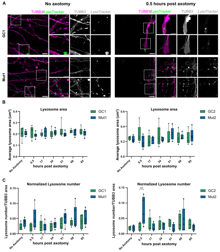
Figure 7.
LRRK2 G2019S is associated with a transient increase in lysosome number in axons proximal to the injury site post axotomy. (A) Representative micrographs of LysoTracker-positive lysosomes in TUBB3-stained axons of GC1 and Mut1 neurons at the distal exit site of microchannels under the no axotomy condition (left) and 0.5 h post axotomy (right). Scale bar: 10 µm, insets 5 µm. (B) Quantification of the average area of lysosomes at different times post axotomy in GC1 and Mut1 neurons (left) and GC2 and Mut2 neurons (right) (C) Quantification of the number of lysosomes normalized to TUBB3-positive area at the distal exit site of microchannels of GC1 and Mut1 (left) and GC2 and Mut2 neurons (right) at different times post axotomy. For all graphs: N = at least five independent experiments. Statistics were calculated using a 2-way ANOVA with Sidak’s multiple comparison test. ** corresponds to p-adj < 0.01.

