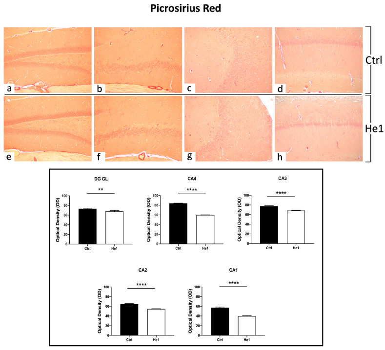
Figure 4.
PSR staining evaluation under light microscopy. Representative hippocampal specimens showing parenchyma and blood vessels from control (a–d) and He1-treated animals (e–h). Microscopy magnification: 20× (a–h). Panel A: Histograms showing OD measurements in DG and CA subregions. p-values calculated by unpaired Student’s t-test. p-values: ** p < 0.01; **** p < 0.0001.

