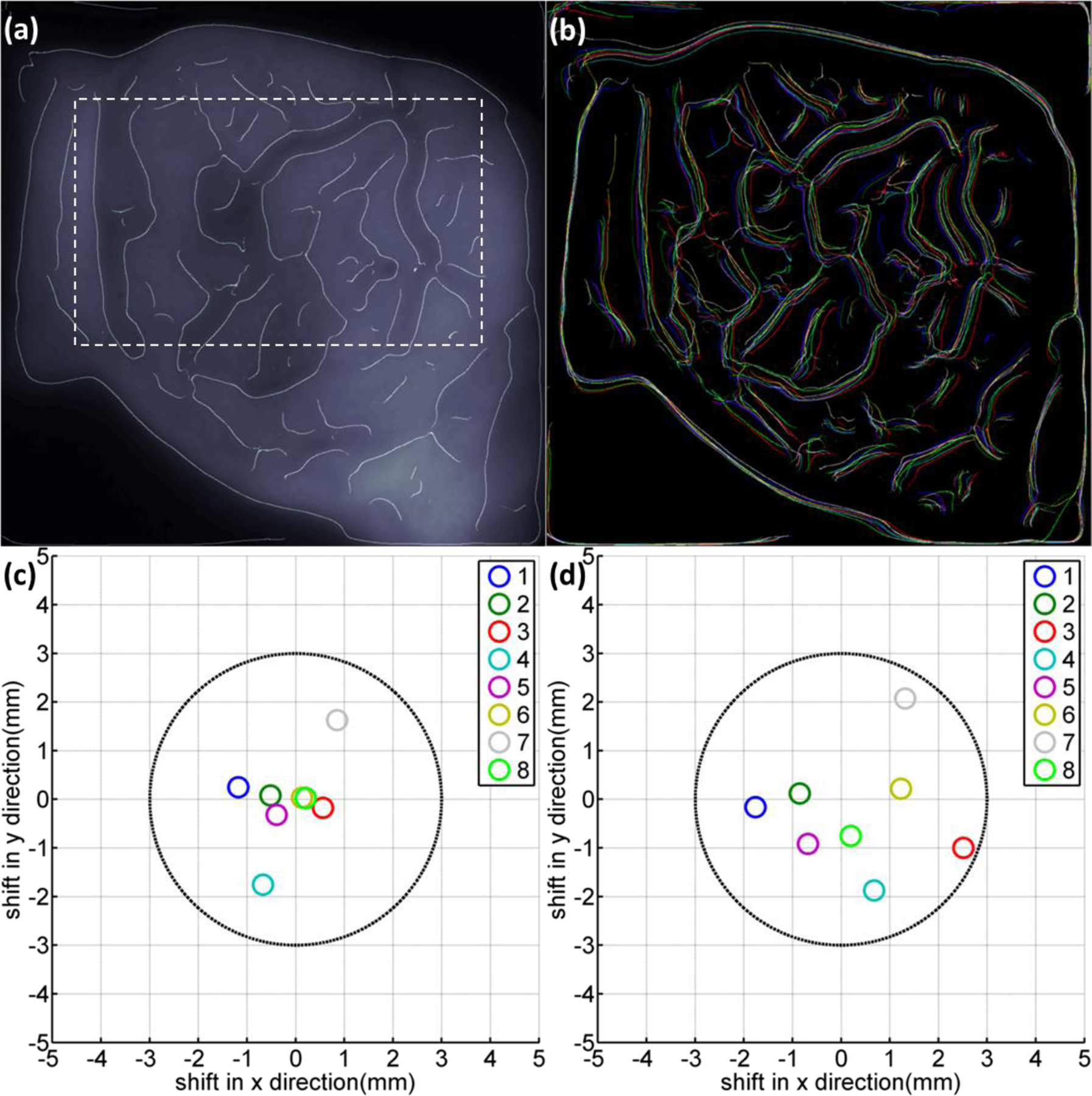
Figure. 4:

(a) The edge enhanced Cherenkov image of patient 3 with a chosen internal region (indicated by dotted lines) within the treatment beam field. (b) Detected edges in Cherenkov images for patient 3, false colored for different imaged treatment fractions. (c) Retrieved positioning errors for patient 3 from rigid image registrations based on the full frame of images. (d) Retrieved positioning errors for patient 3 from rigid image registrations based on the chosen internal region as indicated in (a) with dotted lines.
