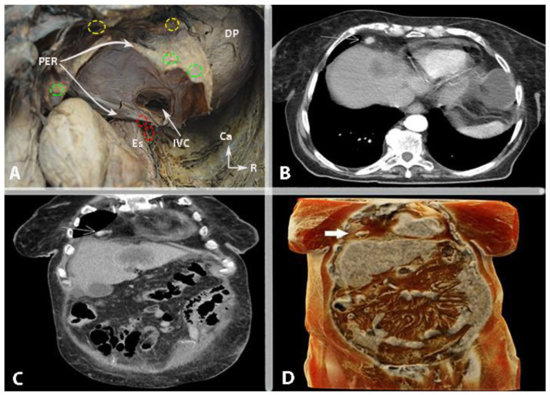
Figure 15.
Cardiophrenic lymph nodes (authors’ own material). (A) Anatomical location of the anterior cardiophrenic lymph nodes (yellow circles—anterior group location; green circles—middle group location; red circles—posterior group location). (B) Contrast-enhanced CT in the axial plane. The arrow points to the metastatic cardiophrenic lymph node. (C) Contrast-enhanced CT in the coronary plane. The arrow indicates the metastatic cardiophrenic lymph node. (D) Three-dimensional CT volume-rendered image. The arrow points to the metastatic cardiophrenic lymph node. PER—pericardium; Es—esophagus; IVC—inferior vena cava; DP—diaphragmatic part of parietal pleura; Ca—caudal, R—right.

