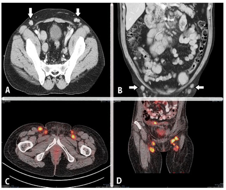
Figure 17.
Imaging of metastatic inguinal lymph nodes in ovarian cancer (authors’ own material). (A) Contrast-enhanced axial CT image of the pelvis. The arrows point to pathologic inguinal lymph nodes. (B) Contrast-enhanced coronal CT image of the pelvis. Arrows point to pathologic inguinal lymph nodes. (C,D) PET/CT—inguinal lymph node metastases.

