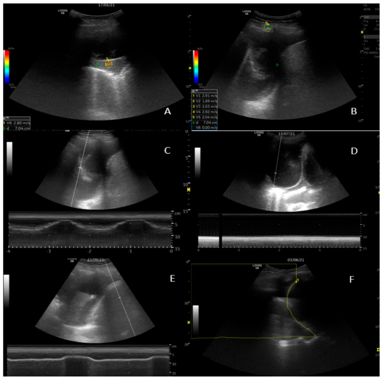
Figure 1.
Ultrasound examinations. (A) 2D shear wave elastography (SWE) of visceral pleura. (B) SWE of parietal pleura with 5 readings recorded in the lower left corner. (C) M-mode measurement of lung movement. The respiratory pattern is seen in the curves on the bottom of the screen. (D) M-mode measurement of lung movement. Here, absence of lung movement is illustrated by the flat line below the 2-D image. (E) M-mode measurement of diaphragm movement. (F) Area-method measurement of area above the diaphragm during inspiration.

