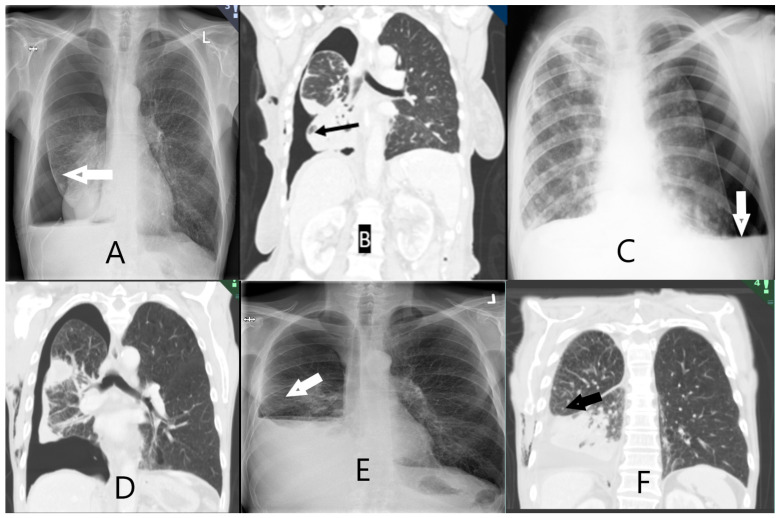
Figure 2.
Radiological images of non-expandable lung (NEL). CXR (image (A,C,E)) and CT-scan (image (B,D,F)). (A) NEL. Lower lobe with thickened visceral lining. (B) NEL. Obstructive tumour of the lower lobe. (C) NEL. Hydropneumothorax following thoracentesis. (D) NEL with thickened pleura. (E) NEL. Small hydropneumothorax following thoracentesis. (F) NEL. Pleural effusion following thoracentesis. Fluid has replaced initial air in the pleural space seen on CXR after thoracentesis.

