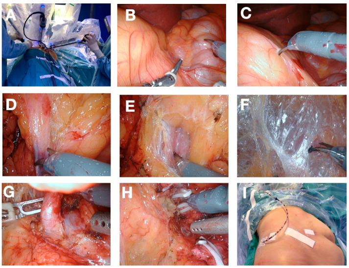
Figure 2.
Description of the procedure showing the intraoperative view of the operation (A) docked robot; (B) view of the anatomy after docking: Colon ascendens (beneath right instrument), kidney shape left to left instrument; (C) incision of the peritoneum between kidney and colon; (D) dissection of the lower pole adherences; (E) view of psoas muscle; (F) avascular plane between psoas muscle and kidney (holding up kidney); (G) Renal artery, (H) dissection of the renal vein after dissection of the renal artery and clamping of the renal vein; (I) final postoperative view after undocking of robot and wound closure.

