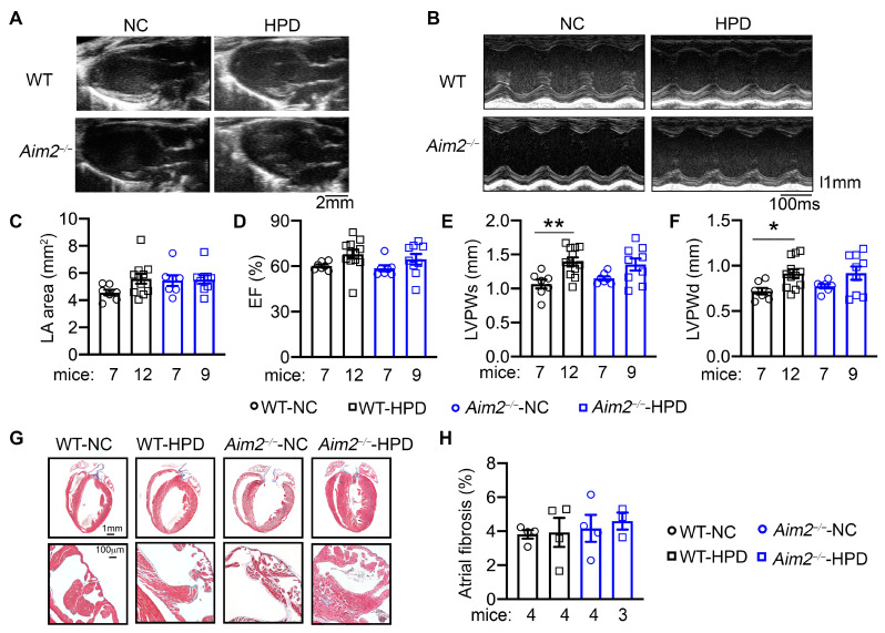
Figure 4.
High-protein diet (HPD) promotes mild ventricular hypertrophy. (A) Representative long-axis echocardiography recording to assess the left atria (LA) area. (B) Representative M-mode echocardiography recordings in four groups of mice at the end of the 4-week feeding period. (C) Quantification of LA area. (D) Quantification of ejection fraction (EF%). (E,F) Quantification of LV posterior wall thickness during systole and diastole (LVPWs, (E); LVPWd, (F)). (G) Representative Masson Trichome’s staining in cardiac tissue. The second row of images indicated the left atria. (H) Quantification of the percentage of fibrosis in atrial tissue. * p < 0.05, ** p < 0.01. p-values were determined by Welch ANOVA and Dunnett’s T3 multiple comparisons test in (E,F).

