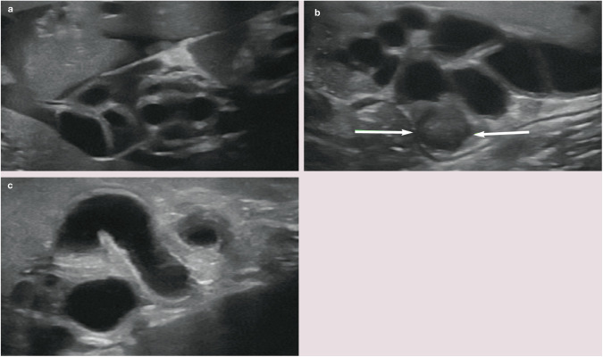
Figure 13.
(a–c) Ultrasound images showing the typical appearance of dilated extrahepatic bile ducts in three cats. Note the relatively thick walls and tortuosity of the ducts. A small volume of echogenic free peritoneal fluid is visible between the hepatic lobes in (a). The arrows in (b) indicate the presence of sludge within the lumen of a dilated extrahepatic duct

