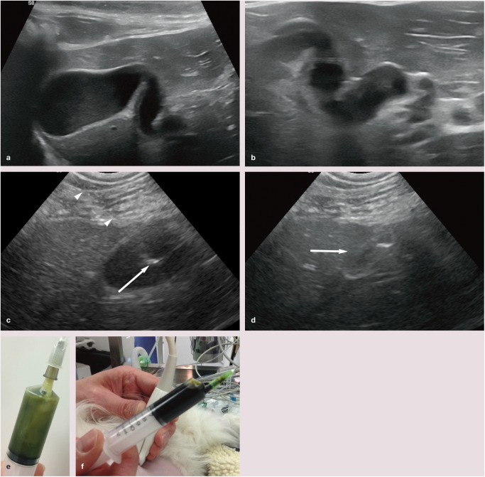
Figure 16.
Images from an 11-year-old female neutered Siamese cat with cholangitis and pancreatitis. (a) On ultrasound, the gall bladder is relatively large and contains non-shadowing sediment (not shown). (b) The wall of the cystic/common bile duct is mildly thickened. The lumen is mildly dilated throughout its length and contains a moderate amount of mobile non-shadowing sludge. (c) Cholecystocentesis was performed with the cat in left lateral recumbency; a right-flank approach is generally the author’s preference. The shaft (arrowheads) of the needle is just visible. The tip of the needle (arrow) is clearly visible within the lumen of the gall bladder. (d) Following aspiration, the gall bladder (arrow) is almost completely empty. The focal hyperechoic structure within the gall bladder is the tip of the needle. (e) The bile from this cat was abnormally pale and turbid. Cytology showed evidence of suppurative inflammation associated with bacteria, consistent with bacterial cholecystitis. Fine-needle aspirates of the liver were performed at the same time and revealed mild hepatocellular lipidosis and evidence of active inflammation. (f) For comparison with (e), this sample acquired following ultrasound-guided cholecystocentesis using a 21 G 1.5 inch needle and 10 ml syringe shows the normal appearance of bile

