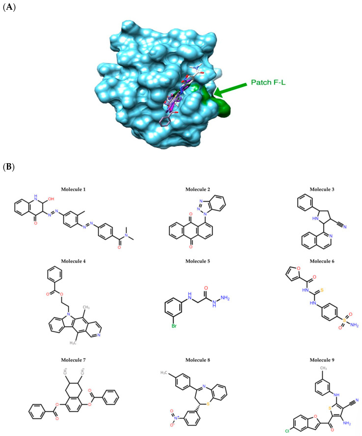
Figure 1.
In silico identification of selective c-FLIP DED-binding molecules. (A) Best docking poses of the nine molecules in the binding pocket of the c-FLIP structural model. The binding pocket is a “hydrophobic patch” of Phenylalanine-Leucine (F-L) motif which is highly conserved in DED domains. The SiteFinder module (MOE) allowed us to identify the druggable pockets. The molecules are in stick representation. c-FLIP structure model is represented as blue surface. The F-L patch is highlighted in green. (B) 2D structures of the nine molecules selected by in silico methods.

