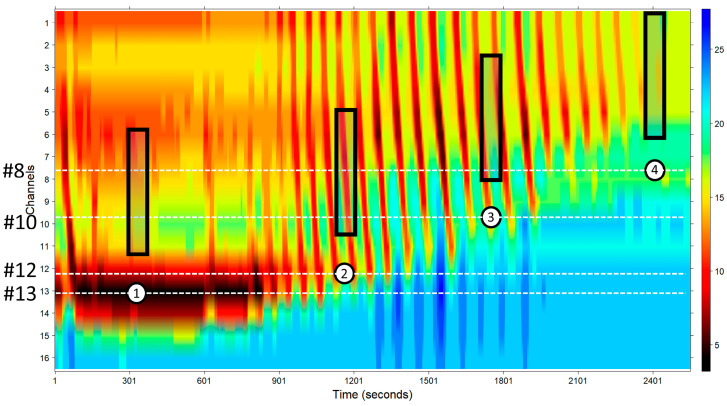
Figure 2.
EndoFLIPTM movements with respect to the esophagus wall. This figure shows how the EndoFLIPTM balloon slides with respect to the esophagus wall. White circles show the EGJ location in several instances in the study. Dotted white lines mark approximately which channels recorded the corresponding diameters. Shadowed black rectangles represent the approximate esophagus area recorded by different sensors.

