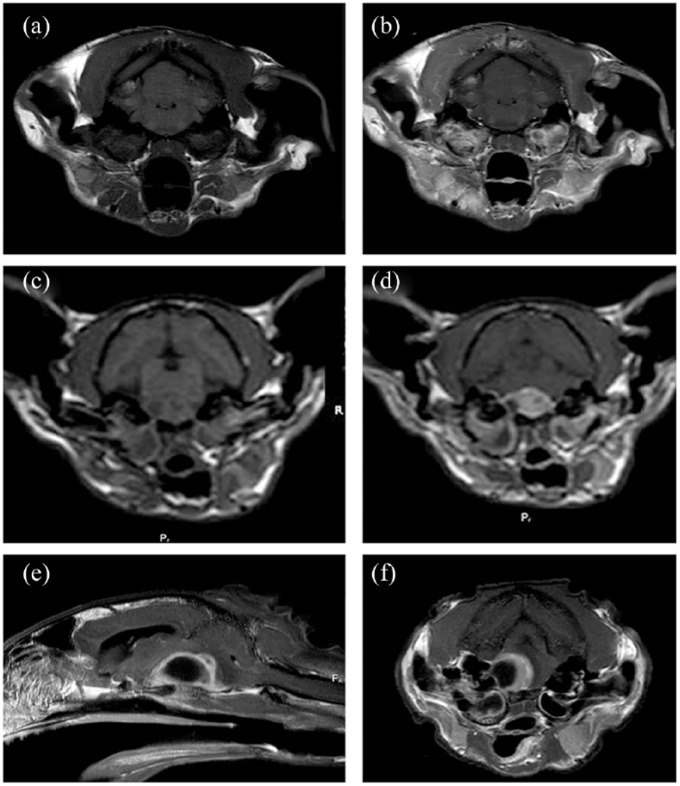
Figure 2.
MRI findings in cats with intracranial complications of otitis media/internal. Significant findings ranged from (a,b) moderate or marked meningeal and/or brainstem parenchymal enhancement adjacent to the petrous temporal bone to (c–f) the presence of a contrast-enhancing mass in the caudal fossa suggestive of an intracranial abscess. (e,f) Note the peripheral contrast enhancement. Panels (a,b) and (d–f) are T1-weighted images acquired after intravenous administration of gadolinium. (c) T1-weighted image acquired prior to administration of gadolinium

