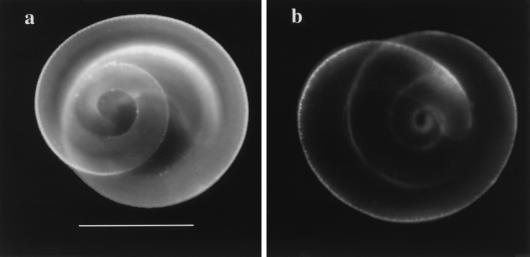
FIG. 3.
Immunofluorescent staining of control (a) and biotinylated (b) larvae. Activated control and biotinylated larvae were prepared as described in the text. Larvae were incubated with 20 μg of MAb 18H per ml before being stained with FITC-conjugated goat anti-rat IgG. Bar, 0.1 mm. The figure was prepared as described for Fig. 1.

