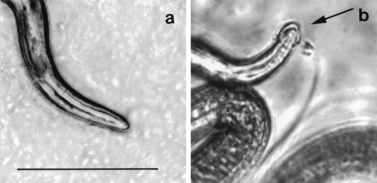
FIG. 4.
Antibody-mediated formation of cephalic caps. Larvae were included in overlays of MDCK cells that were grown to confluence in single-chamber slides and incubated at 37°C under 5% CO2 for 15 min. The images were captured as described in Materials and Methods. (a) Anterior of an infectious larva cultured in medium alone. (b) Cephalic cap on a larva cultured with medium containing 1.0 mg of MAb 18H per ml. Bar, 0.1 mm. The figure was prepared as described for Fig. 1.

