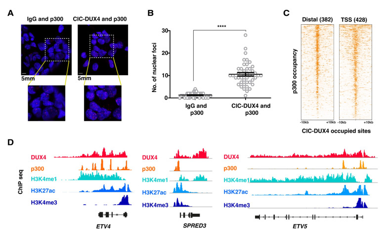
Figure 3.
CIC-DUX4 and p300 proteins interact with each other and share chromatin occupancy: (A) Representative micrographs of PLA signals in the CDS1 cell line, assessed using either DUX4- targeting antibodies (right panels), or isotype-matched IgG (left panels). (B) Dot plot quantification of the number of interaction foci detected in the CDS1 cell lines. Data are presented as mean +/− SD. Statistical analysis was performed using student t-test (quadruple asterisk p < 0.0001). (C) Heatmaps showing the presence of p300 ChIP-seq signals at CIC-DUX4 binding sites in the CDS1 cell line. A 10 kb window centered on both distal (n = 382) and TSS (n = 428) CIC-DUX4 binding sites. (D) Representative ChIP-seq tracks showing chromatin CIC-DUX4 and p300 chromatin co-occupancy as well as H3K4me1, H3K27ac, and H3K4me3 signals at select loci in the CDS1 cell line.

