Figure 5.
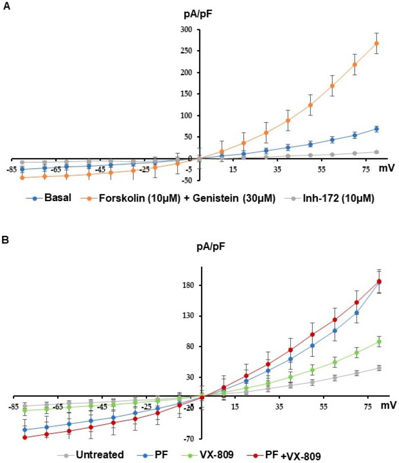
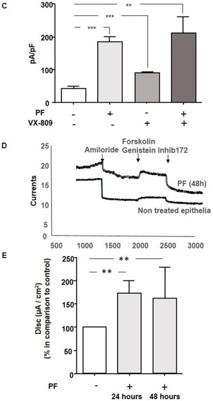
Cl-efflux when MBTP1 is inhibited in native CFBE41o-cells. Cl-efflux via the p.Phe508del-CFTR channel in the presence of PF with or without VX-809 were assessed using patch clamp (whole-cell configuration). (A) Representative I/V curves were obtained in a basal state with CFTR’s activators and inhibitors which were used to assess the specificity of the recorded currents. (B) Representative I/V curves were obtained with PF and/or VX-809. Increased currents via p.Phe508del-CFTR were observed in all conditions when compared to the control condition. The conditions with PF show higher currents than other conditions. (C) The bar graph represents the statistical analysis (Untreated: n = 5; PF: n = 10; VX-809: n = 5; PF plus VX-809: n = 4) of the normalized currents recorded at +80mV. MBTP1 inhibition significantly increases p.Phe508del-CFTR channel currents above more than that of VX-809, but no significant synergistic effect is observed. (D) Example of curves recorded during the Ussing chamber experiments. The upper curve was obtained with PF-treated bronchial epithelial cells from a patient. The lower curve is the recording made with non-treated cells from the same patient. The responses to CFTR’s activators and inhibitor were enhanced with PF. (E) The bar graph represent the statistical analysis of p.Phe508del-CFTR currents recorded on bronchial epithelia from a homozygous p.Phe508del patient in Ussing chamber assays. We show that MBTP1 inhibition (24 and 48 h) significantly increases the p.Phe508del-CFTR currents in comparison to the control condition (n = 3). p < 0.01 (**), and p < 0.001 (***).


