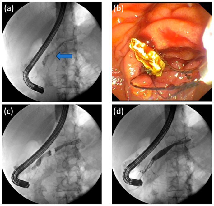
Figure 2.
Endoscopic retrograde cholangiopancreatography is performed using a therapeutic side-viewing duodenoscope (Olympus, Exera TJF 190, Tokyo, Japan). (a) A pancreatogram reveals obstructive filling defect from the head to the body of MPD (as indicated by the blue arrow). (b) Retrieval of the pancreatic duct (PD) calculus using a Trapezoid Basket (Boston Scientific, MA, USA) is depicted. (c) Retrieval of the PD calculus is attempted using balloon extraction (Olympus, Medical Systems, Tokyo, Japan). (d) A pancreatogram reveals complete clearance of the calculus from body and tail of pancreas.

