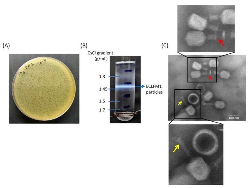
Figure 1.
Morphology and buoyant density of ECLFM1. (A) Plaque morphology on a plate containing bacterial overlay with scale bar of 1 cm. (B) CsCl gradient revealed that the buoyant density of ECLFM1 is 1.45 g/mL. (C) Electron micrograph of ECLFM1 virions with scale bar of 100 nm. The CsCl-purified viral particles were negatively stained with 2% uranyl acetate and visualized at 120K-fold magnification. The phage particle has a constricted tail sheath (yellow arrow) and a tail tube protruding from the tip of the tail (red arrow), which can be seen as a myovirus.

