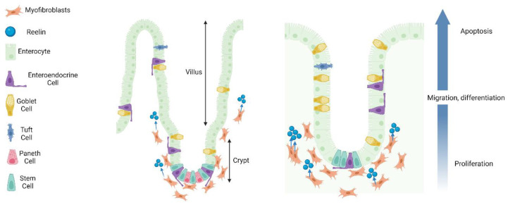
Figure 2.
Crypt–villus axis migration in the small intestine and colon. A schematic delineating crypt–villus axis migration in the small intestine (left panel) and colon (right panel). Reelin is released from subepithelial myofibroblasts to aid in migration processes. Intestinal stem cells of the crypts differentiate into enterocytes, goblet cells, tuft cells, enteroendocrine cells, and Paneth cells (small intestine only). Migrating cells “push” adjacent cells above to encourage cell death and lining regeneration. Additionally, cell death on the villi facilitates proliferative and migratory processes in the crypts. Following migration to villus tips and differentiation, cells live for 3–5 days before being shed into the lumen of the gut. Paneth cells can live for close to 60 days.

