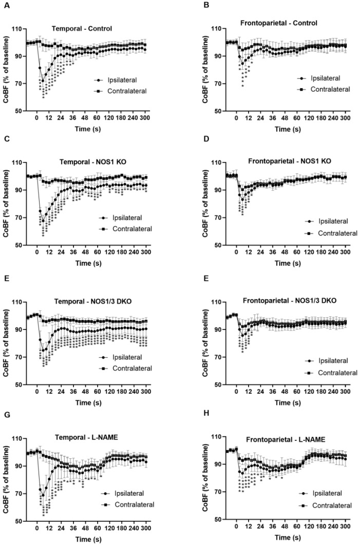
Figure 3.
Cerebrocortical blood flow (CoBF) in the ipsilateral and contralateral hemispheres after unilateral carotid artery occlusion (CAO). Panels A and B: Blood flow changes in temporal (A) and frontoparietal (B) regions in control mice (n = 10) after unilateral CAO. In both ipsilateral regions, the blood flow normalizes after a transient reduction post-CAO. Panels C and D: Blood flow changes in temporal (C) and frontoparietal (D) regions in NOS1 KO animals (n = 6) after unilateral CAO. Note the less complete recovery in the ipsilateral temporal region compared to the frontoparietal region. Panels E and F: Blood flow changes in temporal (E) and frontoparietal (F) regions in NOS1/3 DKO animals (n = 11) after unilateral CAO. Note the sustained hypoperfusion in the temporal region but not in the frontoparietal region. Panels G and H: Blood flow changes in temporal (G) and frontoparietal (H) regions of L-NAME-treated animals (n = 15) after unilateral CAO. Note that acute severe hypoperfusion is both regions of the ipsilateral hemisphere and mild hypoperfusion of the contralateral hemisphere in the first 90 s. after CAO and the recovery of CoBF in all regions thereafter. Values are presented as mean ± SD percentage of the baseline. Circles indicate the ipsilateral, while squares indicate the contralateral hemisphere. Statistical significance is denoted as * p < 0.05, ** p < 0.01, *** p < 0.001, **** p < 0.0001 vs. contralateral (two-way Anova with Bonferroni’s post hoc test).

