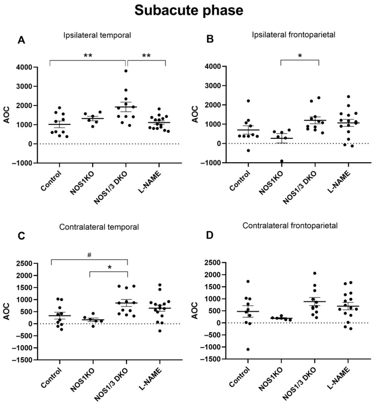
Figure 5.
Comparison of hypoperfusion levels determined by AOC (area over the curve) values of blood flow in control wild type, NOS1 KO, NOS1/3 DKO, and L-NAME-treated wild-type mice in the subacute phase (90–300 s) after unilateral carotid artery occlusion. NOS1/3 DKO animals show worsened recovery in the ipsilateral temporal (Panel (A), ** p < 0.01) and frontoparietal (Panel (B), * p = 0.0400) regions. The blood flow recovery of NOS1/3 DKO mice is also compromised in the temporal (Panel (C), * p = 0.0208 and # p = 0.0514) region, but not in the frontoparietal region (Panel (D)), of the contralateral hemisphere. Scatter dot plots with mean ± SEM are shown. Statistical analysis was performed with one-way Anova and Tukey’s post hoc test).

