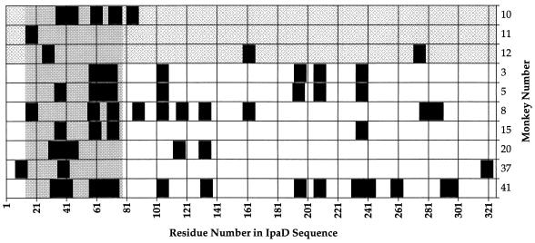
FIG. 2.
Distribution of IpaD peptide epitopes recognized by rhesus monkeys challenged with virulent S. flexneri 2a. The figure shows the peptide epitopes recognized by the convalescent-phase serum samples from monkeys infected with virulent S. flexneri 2a (black rectangles). Peptide epitopes were defined as the peaks with the highest OD405 in the multipin ELISA and were plotted by the residue number of the first amino acid of the peptide as related to the sequence of IpaD. Animals were grouped by symptoms: asymptomatic animals (monkeys 10, 11, and 12) are at the top (grey checkerboard shading), and animals with shigellosis are towards the bottom (no shading). Each of the 10 animals which recognized peptide epitopes on IpaD recognized at least one peptide epitope within the antigenic region between amino acid residues 14 and 77 (solid gray region).

