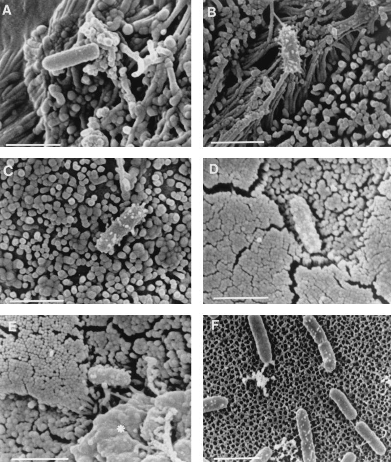
FIG. 1.
Scanning electron micrographs of S. typhimurium SL1344 adhering to Caco-2 cells (A and B), MDCK cells (C), murine Peyer’s patches (D and E) and cell-free culture supports (F). A proportion of wild-type S. typhimurium cells adhering to target cells formed numerous short filamentous appendages when they adhered to either Caco-2 cells (B) or MDCK cells (C), while others lacked such appendages (A). Similar appendages were present on some S. typhimurium SL1344 cells that adhered to M cells (D and E) after infection of murine Peyer’s patches in ligated intestinal loops. The asterisk in panel E indicates an area of membrane remodelling associated with bacterial invasion. A small proportion of S. typhimurium SL1344 cells attached to cell-free culture supports possessed appendages which exhibited a similar distribution but which were shorter than many of those on cell-adhered bacteria (F). Bars, 2 μm.

