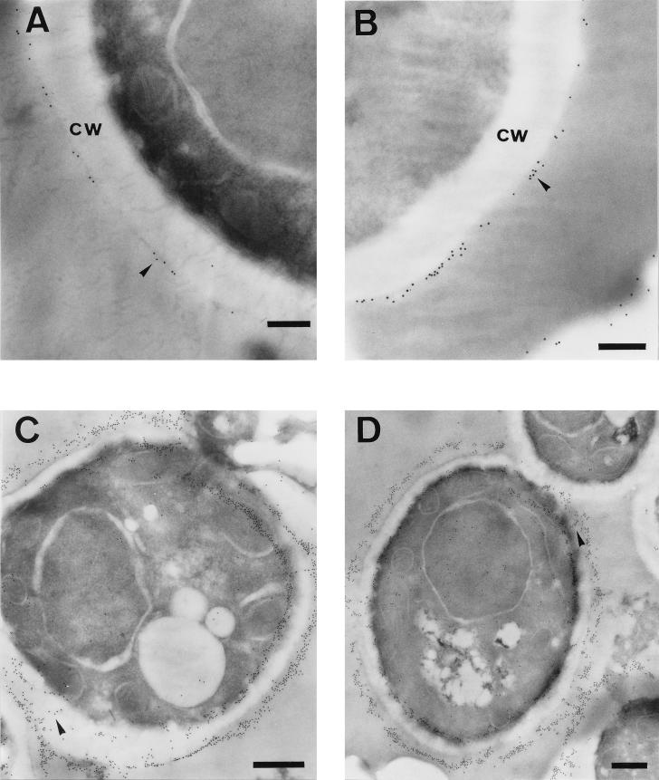
FIG. 1.
Immunoelectron microscopy detection of GAPDH protein in C. albicans yeast cells by the preembedding (A and B) and postembedding (C and D) methods. The labelling was detected at the outermost layer of the cell wall (cw) in a patchy distribution (A and B, arrowheads). The antigen also appears extending through the cell wall (C and D, arrowheads). The label was also detected in the cytoplasm (C and D). Bars, 0.25 μm (A and B) and 0.5 μm (C and D).

