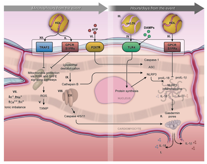
Figure 1.
Minutes/hours from MI and hours/days from MI. The NLRP3 inflammasome cellular mechanism initiates during the first minutes of MI and can persist for days, exerting an influence on acute or chronic outcomes. Activation of pro-IL-1β and pro-IL18 and their respective release outside the membrane (I); gasdermin-D on the lytic cell death due to the pore formation on the cellular membrane, characterized by proinflammatory cell death or pyroptosis (II); proinflammatory oxidized LDL (III); TLR4 and NLRP3 inflammasome transcription pathway (IV); MI exacerbates the production of mitochondrial ROS in a process mediated by the TXNIP (V); P2X7R receptor under ATP stimulation promotes calcium and sodium influx, resulting in potassium efflux and classic signaling for NLRP3 inflammasome activation (VI); ionic imbalance (mainly by increase of Ca2+ mobilization and decrease of K+ efflux) (VII); endoplasmic reticulum stress, triggering caspase 4/5/11 activation (VIII); Cathepsin B, inducing NLRP3 activation (VIX); S1PRs play a crucial role in the interaction of S1P, facilitating cytoprotective mechanisms both short- and long-term, following MI (X and XI). The activation of the Tumor Necrosis Factor 2 Receptor (TRAF2) by HDL can influence the activity of sphingosine kinase-1 and consequently increase the production of sphingosine-1-phosphate (XII). The arrows mean the increase (↑) or decrease (↓) in ion concentration into the cytoplasmic milieu.

