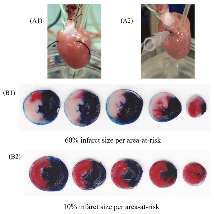
Figure 2.
Representative image of isolated perfused hearts (ex vivo) using Langendorff apparatus undergoing ischemia and reperfusion protocol. Rat heart during regional ischemia (A1) and reperfusion (A2). Figures represent groups that received vehicle (B1) or HDL (200 µg/mL) infusion (B2), after the protocol of staining with 2,3,5-triphenyltetrazolium chloride staining. The heart sections (B1,B2) are presented in tree colors for histologic analysis: blue, the non-ischemic area (also known as non-risk area); red, the ischemic area (also known as area-at-risk); and white or pale, the infarct area. The infarct size was calculated as a percentage of the area-at-risk.

