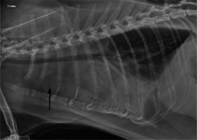
Figure 4.

Lateral thoracic radiograph from case 2 presented for respiratory distress. Note the pleural effusion (most evident cranioventrally) and prominent sternal lymph node (arrow). Although this is an atypical presentation, it is important to emphasise that some cases of FGESF can present with bicavitary involvement, the disease process starting in the abdomen and spreading to the thoracic cavity, presumably due to drainage of abdominal lymphatics to the sternal lymph node. A similar observation was reported recently3
