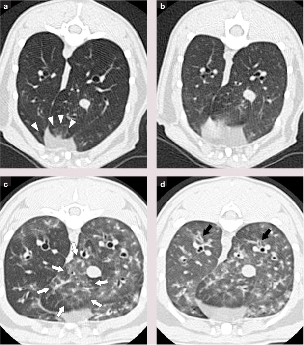
Figure 3.
Thin section CT images in cats with experimentally induced asthma, where white arrows and arrowheads show areas of increased lung attenuation and black arrows indicate areas of bronchial wall thickening: (a) MSC-treated cat during inhalation; (b) MSC-treated cat during exhalation; (c) untreated cat during inhalation; (d) untreated cat during exhalation.31 Airway remodeling, as represented by lung attenuation and bronchial wall thickening scores, was significantly decreased in MSC-treated cats at 9 months after five intravenous injections of allogeneic adipose-derived MSCs

