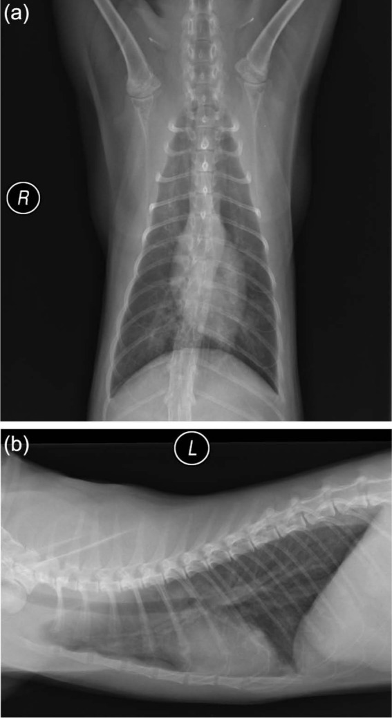
Figure 1.

Ventrodorsal (a) and lateral (b) thoracic radiographs of a cat (EXP-Aa) taken 148 days after experimental infection with 51 Aelurostrongylus abstrusus larvae. The radiographs reveal a patchy, moderately increased soft tissue opacity in the lungs with a predominantly bronchocentric distribution. R = right; L = left lateral
