Figure 2.
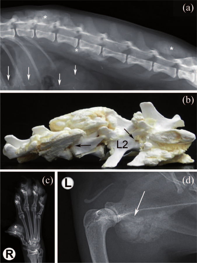
(a) Lateral view of the T11–S1 spine. Amorphous calcified masses superimposed on vertebral arches and spinous processes of the T13–L3 and L6–L7 spinal segments, and transverse process of L3 are visible (asterisks). Linear mineral opacities within the cranial abdomen are visible (arrows). (b) Gross evaluation of the T12–L3 spinal segment; multifocal calcified masses stenosing lateral foramina (arrows), and involving L1–L2 spinous processes and articular facets. (c) Dorsoventral radiograph of the right forepaw. The amorphous, calcified mass along the middle and distal phalanges of the first toe is well appreciable (asterisk). (d) Mediolateral view of the left scapulo-humeral joint. Amorphous calcified mass arising from the scapula and superimposing on the joint is clearly visible (arrow). R = right; L = left

