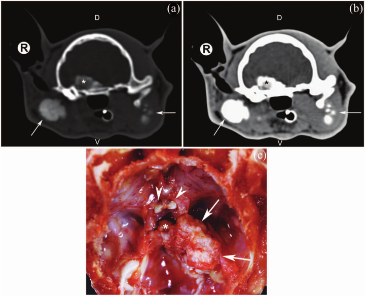
Figure 3.
Pre-contrast bone window (WW = 2000 HU; WL = 350 HU) (a) and post-contrast soft tissue window (WW = 360 HU; WL = 40 HU) (b) transverse computed tomographic images of the skull of the same cat as in (a). A non-vascularised, amorphous, mineralised mass, measuring 13.6 × 13 × 10.3 mm, originating from the right lateral skull base and superimposing on the sella was revealed (asterisk). Images were obtained with a third-generation CT and a 1.5 mm slice thickness. Arrows indicate mineralised masses lateral to the right tympanic bulla and left mandibular ramus. (c) Dorsal view of the basisphenoid bone after brain removal of the same cat as in (a); mineralised mass (arrows), optic nerves (arrow heads) and hypophisis (asterisk). R = right; V = ventral; D = dorsal; WW = window width; WL = window level

