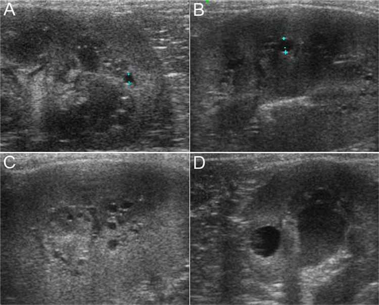
Figure 1.
Ultrasonographical images of the kidneys of Maine Coons with renal cysts. (A) Patient 6, sagittal view of the caudal pole of the right kidney. Between the calipers, a 1.8 mm cyst at the corticomedullary junction. Fine hyperechoic dots are aligned along the deeper portions of the medulla (rim sign). Renal parenchyma was interpreted as otherwise normal. Sagittal (B) and parasagittal (C) views of the left kidney of patient 3. Between the calipers is a 3 mm cyst. Countless minuscule cysts are visualised along the corticomedullary junction. (D) Patient 5, transverse view of the right kidney. The largest cyst observed in this series (5.9 mm in diameter) is found within the cortex

