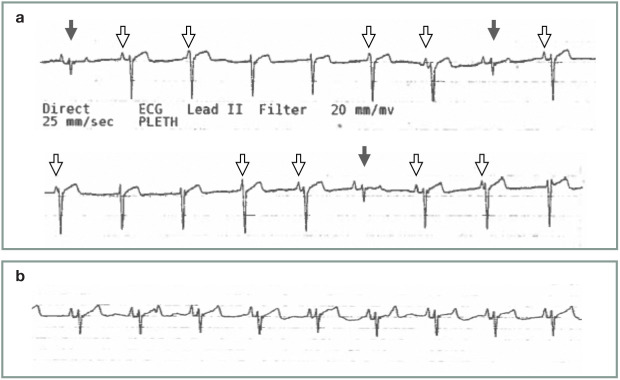
Figure 25.
(a) ECG tracing from an anesthetized cat demonstrating synchronous atrioventricular dissociation. Note the largely negative QRS complexes that are junctional in origin, as well as the smaller, normally conducted QRS complexes (solid arrows). The P waves (open arrows) are seen to be masked by junctional escape beats, indicating a slightly faster rate of junctional depolarization as compared with sinus node depolarization. (b) ECG tracing from the same patient after administration of an anticholinergic agent (glycopyrrolate 0.01 mg/kg IV). Note the normal sinus rhythm, with slightly increased rate of sinus node depolarization and lack of junctional escape beats. ECG tracings in (a) and (b) were recorded at a paper speed of 25 mm/s. Courtesy of Gregg Griffenhagen

