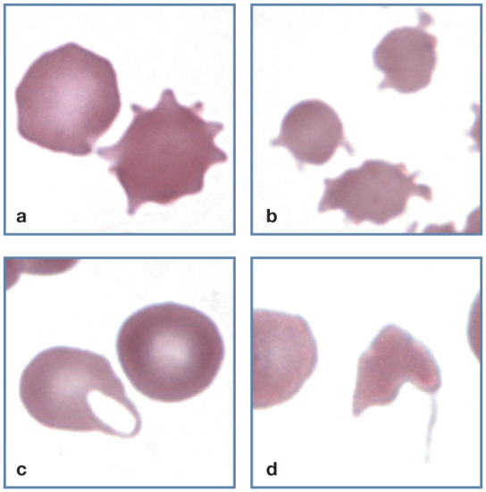
Figure 10.

Abnormal erythrocyte shapes. (a) A normal discocyte (left) and an echinocyte (right) in blood from a cat with uremia. (b) Three acanthocytes in blood from a cat with hepatic lipidosis. (c) A keratocyte with intact vacuole (left) and a discocyte (right) in blood from a cat with a portosystemic shunt. (d) A schistocyte in blood from a cat with diabetes mellitus, interstitial lung disease and erythrocyte fragmentation. Wright-Giemsa stain
