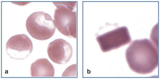
Figure 12.

Abnormal erythrocyte shapes. (a) Three eccentrocytes in blood from a cat with acetaminophen toxicity. Pale Heinz bodies are also visible in two of the eccentrocytes (left and top). (b) A hemoglobin crystal is present within an erythrocyte. The faintly stained cell membrane (top) is visible. Wright-Giemsa stain
